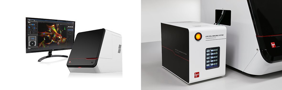
CELENA® X High Content Imaging System (616)
The CELENA® X High Content Imaging System is an integrated imaging system designed for rapid, high content image acquisition and analysis. Customizable imaging protocols, image-based and laser autofocusing modules, and a motorized XYZ stage simplify well plate imaging and slide scanning. The integrated CELENA® X Cell Analyzer software processes images and data for quantitative analysis. Analysis pipelines can be put together and reused to identify cellular or subcellular objects, process images for optimal data collection, and make various measurements.
Features:
Temperature and CO2 control
Laser AutoFocus module and image-based autofocus
Phase contrast condenser
Motorised xyz stage for scanning well plates and slides
Long-term time-lapse acquisition (multiple days)
Objectives:
Olympus 4x/0.13 NA air, WD=17 mm
Olympus 10x/0.3 NA air, WD=10 mm
Olympus 20x/0.45 NA air, WD=6.60-7.70 mm
Olympus 40x/0.6 NA air, WD=2.70-4.00 mm
Filter cubes:
DAPI (Ex 375/28, Em 460/50)
EGFP (Ex 470/30, Em 530/50)
RFP (Ex 530/40, Em 605/55)
Cy5 (Ex 620/60, Em 700/75)
Guides
Book an instrument or training:
Contact
Microscopy Facility Manager
Rumelo Amor
r.amor@uq.edu.au
Senior Microscopy Officer
Andrew Thompson
a.thompson4@uq.edu.au
Senior Microscopy Officer
Zac Pujic
z.pujic@uq.edu.au
Histology Facility Manager
Rob Sullivan
qbi.histology@uq.edu.au
To contact the microscopy team please use:
qbi.microscopy@uq.edu.au