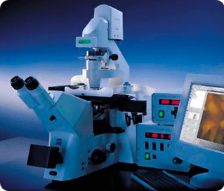
Epifluorescence Microscopes: Axio Observer Indigo (616) - Inverted
Inverted fluorescent microscope fitted with a monochrome camera. Suited for fluorescence and bright field imaging of live cells in flasks, dishes or glass-bottomed MatTek dishes.
Features Include:
- CO2 and temperature incubation controls for live cell imaging
- Fully motorised X-Y-Z stage
- Z-stack imaging
- MosaiX tiled imaging
- Mark and Find for fully automated image capture and high-throughput experiments
- ApoTome optical sectioning
- Objectives have long working distances (good for plastic dishes)
- Different stage inserts for small dishes, multi-well plates and flasks
- Heated stage inserts for MatTek dishes and 24well plates
- DIC
- plasDIC for DIC imaging through plastic
Objectives:
- Air:
- 2.5x 0.16 NA / 18.5mm WD / 2.58 μm/pixel,
- 10x 0.3 NA / 5.2mm WD / 0.645 μm/pixel,
- 20x 0.8 NA / 0.55mm WD / 0.323 μm/pixel,
- 20x 0.4 NA / 7.9mm WD / 0.323 μm/pixel,
- 40x 0.6 NA / 2.9mm WD / 0.161 μm/pixel
- Glycerine:
- 63x 1.3 NA / 0.17mm WD / 0.102 μm/pixel
Fluorescent Filter Sets For:
- DAPI (FS#49) BP335-383/BS395/BP 420-470
- GFP / Alexa 488 (FS#38) BP450-490/BS495/BP500-550
- Cy3 / Alexa 546/555/568 (FS#43) BP533-558/BS570/BP570-640
- mCherry / Alexa 594 (FS#43) BP533-558/BS570/BP570-640
- Cy5 / Alexa 633/647 (FS#50) BP625-655/BS660/BP665-715
Fluorescent Illumination:
- Colibri LEDs – 365nm (DAPI), 470nm (GFP), 530nm (Alexa546/Alexa555/Cy3/mCherry), 625nm (Cy5/Alexa647)
Instructions
Guide to using Axio Imagers and Observers
Guide to High-throughput Imaging
When including this microscope in your methods:
Cells/tissue/specimens were imaged on an Axio Observer Z1 inverted fluorescence microscope (Carl Zeiss Pty Ltd) fitted with a full-enclosure incubator with CO2 and temperature control for live-cell imaging, an Axiocam 702 monochrome high-speed camera (Carl Zeiss), and a 20x/0.8 NA Plan-Apochromat objective (or other – please refer to objectives list) (Carl Zeiss). Image acquisition was performed using ZEN software (Carl Zeiss).
