Epifluorescence Microscopes: Axio Imager Green (414)
Upright fluorescent microscope fitted with both monochrome and colour cameras. Suited for fluorescence and bright field imaging of fixed samples on slides.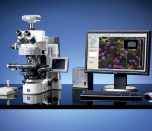
Features Include:
- Fully motorised X-Y-Z stage
- Z-stacks
- Mosaix tiling
- Mark and Find
- Fully automated image capture experiments
- ApoTome optical sectioning
- DIC
Objectives:
- Air:
- 5x 0.16 NA / 18.5mm WD / 1.29 μm/pixel,
- 10x 0.45 NA / 2mm WD / 0.645 μm/pixel,
- 20x 0.8 NA / 0.55mm WD / 0.323 μm/pixel,
- 40x 0.75 NA / 0.71mm WD /0.161 μm/pixel
- Oil:
- 63x 1.4 NA / 0.19mm WD / 0.102 μm/pixel
- 100x 1.3 NA / 0.2mm WD / 0.065 μm/pixel
Fluorescent Filter Sets For:
- DAPI (FS#49) BP335-383/BS395/BP 420-470
- GFP / Alexa 488 (FS#44) BP455-495/BS500/BP505-555
- Cy3 / Alexa 546/555/568 (FS#43) BP533-558/BS570/BP570-640
- DAPI / BFP / GFP / Alexa 488 / HcRed / mCherry / Alexa 594 (FS#62HE)
- BP450-390/BS395/BP402-448
- BP460-488/BS495/BP500-557
- BP567-602/BS610/LP615
- Cy5 / Alexa 633/647 (FS#50) BP625-655/BS660/BP665-715
- CFP (FS#47) BP426-446/BS455/ BP460-500
- YFP (FS#46) BP490-510/BS515/BP520-550
Fluorescent Illumination:
- CoolLED pE-300 LEDs – 365nm (DAPI), 460nm (GFP/Alexa488), GYR (525-660nm) (Alexa546/555/568/594/mCherry/Cy5/Alexa647)
Instructions
Guide to using Axio Imagers and Observers
When including this microscope in your methods:
Cells/tissue/specimens were imaged on an Axio Imager Z1 upright fluorescence microscope (Carl Zeiss Pty Ltd) fitted with an Axiocam 506 mono(or 512 colour) camera (Carl Zeiss), and a 20x/0.8 NA Plan-Apochromat objective (or other – please refer to objectives list) (Carl Zeiss). Image acquisition was performed using ZEN software (Carl Zeiss).