Diagnosing concussion with image tests
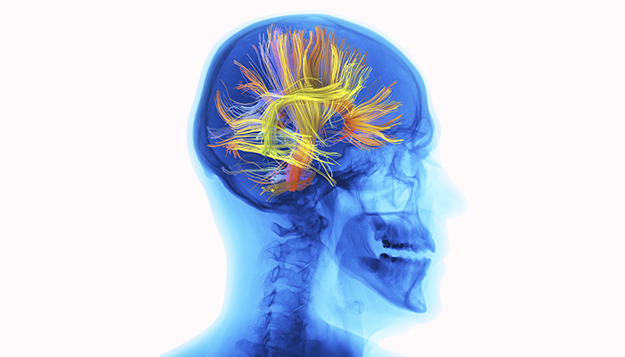
Visualising concussion-related changes in the brain is particularly challenging. Unlike the bleeding inside the brain that might occur with a severe head impact, no obvious structural changes accompany concussion. This lack of obvious damage means that much of the imaging research in concussion focuses on changes in brain function rather than brain structure.
Diagnosing concussion with MRI
Although techniques such as magnetic resonance imaging (MRI) are usually unable to detect structural changes in the brain following a concussion, specific specialised MRI scans can. MRI uses a strong magnetic field and radio waves to build up a picture of the brain. One imaging technique that can currently detect structural changes in the concussed brain is diffusion imaging, which looks at patterns of water movement through brain tissues.
Diffusion imaging can reveal some of the subtle damage of concussion, icluding, for example, evidence of disruptions in the white matter. White matter is like the ‘skeleton’ of the brain; it’s a framework made up of the main parts of nerve cells – the long sections known as axons – and glial cells, which are the most common type of cells throughout the central nervous system.
These disruptions to the white matter are microscopic breaks that, Professor Reutens explains, may affect connectivity and impair brain function. Instead of neurons firing together in coordinated networks, they are out of sync.
Concussion detected using MR spectroscopy
A second approach to studying concussion using imaging technology involves a technique called magnetic resonance (MR) spectroscopy, which looks at changes in brain chemistry. MR spectroscopy is revealing that certain substances produced by brain cells as part of their usual activity are altered by concussion. This is the case even in people who don’t show outward signs of concussion. This suggests that in even the mildest concussion, the energy processes going on in brain cells are altered by the impact.
Impacts of concussion shown by fMRI
Researchers can look at changes in brain function by using another type of MRI known as a functional MRI (fMRI). This imaging allows us to see regions in the brain that are active. QBI's Dr Fatima Nasrallah has been using fMRI and other imaging techniques to study what happens to the brain in the immediate aftermath of a concussion, as well as in the following weeks and months.
Specifically, she has been examining brain networks, which are the different patterns of activity associated with different brain functions. There are networks in the brain that are engaged during different activities, for example, during movement or emotional responses. The symptoms in diseases such as Alzheimer’s disease and schizophrenia result from disruption to these networks.
For concussion, research is showing a similar brain network disruption, but one network in particular is affected. It’s known as the ‘default mode’ network and it’s the one that’s most active when our brain is in what could be called ‘idling mode’. We don’t yet understand why this network is so important, or why it is particularly badly affected by concussion. But, says Dr Nasrallah, changes to this network are apparent soon after concussion.
Imaging techniques useful for diagnosing concussion
Brain imaging studies are helping us understand what is going on in the brain during concussion. It’s also hoped they might show characteristic changes with concussion that can be used to diagnose these conditions and monitor recovery. The reality is that what researchers are likely to find are a series of indicators that can be used by doctors in combination with clinical assessments to make a diagnosis of concussion and assess the recovery time required. That will enable doctors to predict who is more likely to suffer long-term consequences from concussion and allow them to intervene early to prevent or limit the damage.
![]()
![]()
![]()
![]()
![]()
Help concussion research
QBI newsletters
Dr Fatima Nasrallah has a very personal reason for dedicating her professional life to the study of brain trauma. Her grandmother had dementia and Parkinson’s disease, which worsened dramatically after a fall. Since then, Dr Nasrallah has applied the full spectrum of imaging technologies to advance our understanding of what is going on inside the brain during and after a traumatic brain injury. She began her research career studying biochemical brain changes using imaging, then shifted her focus to use MRI to study physical brain changes in animals. A move to a clinical research facility in Singapore brought Dr Nasrallah into contact with military personnel who had been exposed to blast injuries. It allowed her to begin exploring how these injuries could be visualised using imaging technologies. Dr Nasrallah is now leading a large QBI concussion study that will follow people with concussion over a long period of time. She is using imaging technologies to study brain changes and the effects of different treatments and interventions to reduce long-term damage from concussion. |