How to measure brain activity in people
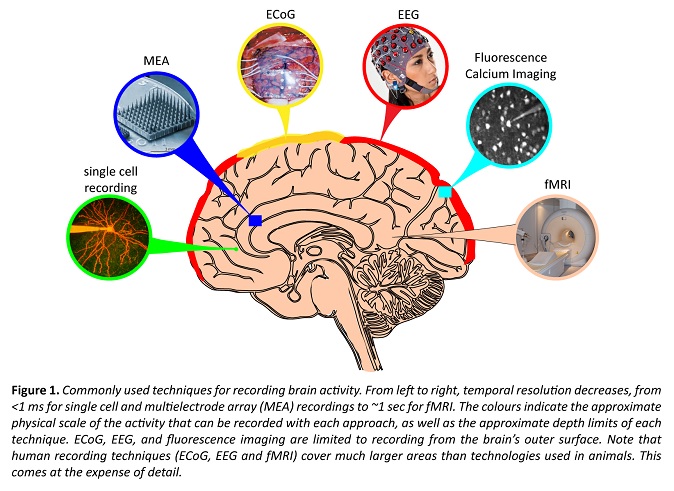
The brain is difficult to study not only because of its inherent complexity; the billions of neurons, the hundreds or thousands of types of neurons, the trillions of connections. The brain also works at a number of different scales, both in the physical sense and in the time domain.
To understand the brain’s electrical activity at these scales, no single technology is enough. As a result, neuroscientists have a suite of tools at their disposal. Some of these, such as fMRI and EEG, can be used in humans because they are non-invasive; they work through by looking into the skull.
But these tools suffer from a lack of detail. To get a more microscopic picture of neuron activity, researchers turn to animal models. This allows the behaviour of individual neurons, or small groups of neurons, to be analysed in much greater detail.
Functional Magnetic Resonance Imaging (fMRI)
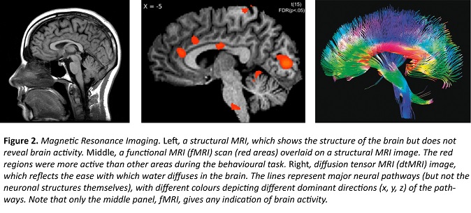
Functional magnetic resonance imaging, or fMRI, might be the most widely known technology for recording neural activity, but it doesn’t actually record activity of neurons – instead, the multicolour images you see of particular brain regions being lit up reflect blood flow in the brain. More precisely, the signal you see reflects the relative presence of oxygenated versus deoxygenated blood; active regions require more oxygenated blood, and so despite being indirect, fMRI allows scientists to infer activity patterns of neurons.
fMRI has become a staple of modern neuroscience research because it allows brain anatomy (obtained from a structural, rather than functional, MRI scan) and function to be correlated in humans. But it does have limitations. Both the spatial (~1 mm3, relating to location) and temporal (~1–2 sec, relating to time) resolutions are poor compared to what we’d want; a cubic millimetre contains around 60,000 neurons – enough to sustain the entire life of a fruit fly or lobster – and complex perceptual decisions take only hundreds of milliseconds, but fMRI provides no access to this information.
Nevertheless, fMRI allows an unrivalled look at where and to what extent different functions may be localised within the human brain, and researchers continue to devise ways to improve its spatial and temporal resolution, for example by making the technique sensitive to neuronal changes rather than changes in blood flow. No current technique matches fMRI for its ability to ‘map’, or determine the likely source of, cognitive function within the human brain.
Electroencephalography (EEG)
Electroencephalography, or EEG, is probably the second-best known technique for recording neural activity. Whereas fMRI records blood flow, a proxy of neuron activation, EEG directly records the brain’s electrical activity via electrodes placed on the scalp of the subject.
However, EEG doesn’t record action potentials, the electrical events that neurons use to communicate with each other. Instead, it surveys the summed activity of hundreds of thousands or millions of neurons in the form of oscillatory activity. In contrast to action potentials, it’s not known what information these oscillations actually carry, but different frequencies of oscillation do correlate with different behavioural states.
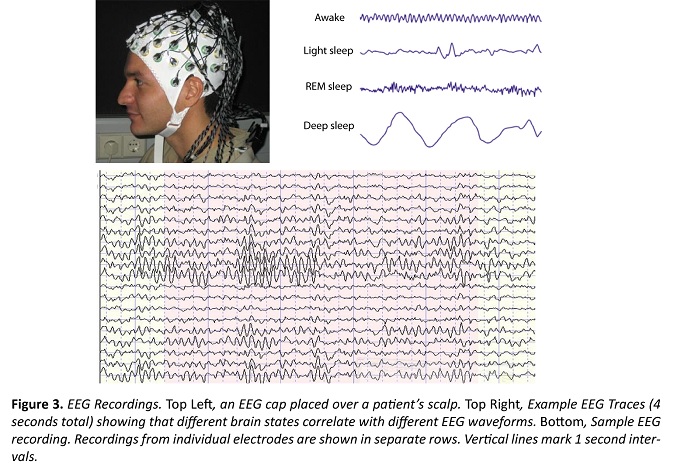
EEG has a 'temporal resolution' far superior to fMRI (~1 ms vs. 1 sec). Because of this, EEG can be used to more accurately track neural dynamics in awake humans, and is often used to determine the brain’s electrical response to a stimulus or condition.
The primary limitation of EEG is its poor spatial resolution, much poorer than for fMRI. Although it’s known that EEG signals only come from the cerebral cortex, it remains extremely difficult to know precisely where in the cortex signals arise.
Furthermore, its cortical bias means that we can’t use it to measure what happens in the hippocampus, where many memories are made and stored, or in the substantia nigra or striatum, regions affected by Parkinson’s Disease. So unlike fMRI, mapping of activity is not really possible with EEG.
Electrocorticography (ECoG)
Electrocorticography is similar to EEG in that it measures the combined activity of millions of neurons, often in the form of oscillatory waves. But there are two major differences. First, ECoG requires insertion of the electrode array under the scalp, and so requires surgery. For this reason, ECoG is only suitable for patients already scheduled for a medical surgery that involves opening of the scalp.
Second, ECoG allows significantly improved localisation of the activity source, as well as the recording of higher frequency electrical activity. Both of these characteristics assist during epilepsy surgery, but for pure research purposes, the technique is too invasive to be used in humans who don’t already require brain surgery.
Summary
Neuroscientists are justifiably limited in the sort of approaches they can use to study human brain activity. However, so far no technology exists that allows detailed neuron activity to be recorded through the human skull, meaning that the measures we can take give fairly coarse information as to how our brains work. These spatial and temporal resolution limits will undoubtedly be improved in the near future, enabling more precise measurements and greater insights into human brain activity. Furthermore, complementary approaches that allow the temporary disruption of neuronal processing will help us to understand what sorts of regional brain dysfunction might lead to the cognitive deficits associated with mental disorders.