Studying synaptic plasticity and learning
Using electrical responses to track plasticity
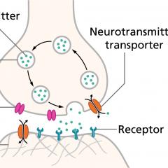
One method to study plasticity is to avoid trying to visualise synapses, and instead to infer what is happening by measuring the electrical currents that they produce. The strength of a synapse depends on how much electrical current it passes, and there are various ways of measuring this. Measuring the electrical response of neurons is the most common means of tracking synaptic plasticity.
Calcium imaging
Another method, one that uses advanced microscopes to visualise living synapses, is to measure how much calcium the postsynaptic neuron receives. Calcium is an extremely important messenger in neurons, and at synapses, the amount of calcium that enters dendritic spines can serve as a proxy for synaptic strength. Researchers can use fluorescent dyes to track synaptic activity and plasticity. The advantage of fluorescent calcium imaging over electrical recordings is that plasticity can be monitored at individual synapses, rather than at the level of the entire neuron.
Playing hide-and-seek with synapses
Researchers can also look at the appearance and disappearance of synapses. Not only can synapses become stronger and weaker with experience (LTP and LTD), but new ones can form and others can be lost. These synaptic changes can be tracked by imaging individual dendritic spines in live animals, over periods of days or weeks, as the animal learns.
In summary, synaptic plasticity is undoubtedly one of the most important phenomena in neuroscience. What makes the brain amazing is that it changes with experience, allowing us to learn from and adapt to the world around us. Synaptic plasticity is a very large part of this, although neurogenesis is increasingly viewed as another vital contributor, at least in certain parts of the mature brain.