What is the blood-brain barrier?
The brain is precious, and evolution has gone to great lengths to protect it from damage. The most obvious is our 7mm thick skull, but the brain is also surrounded by protective fluid (cerebrospinal – of the brain and spine) and a protective membrane called the meninges. Both provide further defence against physical injury. ![]()
Another protective element is the blood–brain barrier. As the name suggests, this is a barrier between the brain’s blood vessels (capillaries) and the cells and other components that make up brain tissue. Whereas the skull, meninges and cerebrospinal fluid protect against physical damage, the blood–brain barrier provides a defence against disease-causing pathogens and toxins that may be present in our blood.
The blood–brain barrier was discovered in the late 19th century, when the German physician Paul Ehrlich injected a dye into the bloodstream of a mouse. To his surprise, the dye infiltrated all tissues except the brain and spinal cord. While this showed that a barrier existed between brain and blood, it wasn’t until the 1960s researchers could use microscopes powerful enough to determine the physical layer of the blood–brain barrier.
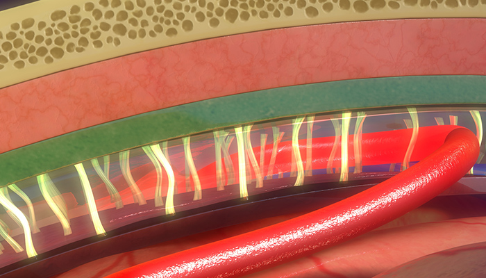
We now know the key structure of the blood–brain barrier that offers a barrier is the “endothelial tight junction”. Endothelial cells line the interior of all blood vessels. In the capillaries that form the blood–brain barrier, endothelial cells are wedged extremely close to each other, forming so-called tight junctions.
The tight gap allows only small molecules, fat-soluble molecules, and some gases to pass freely through the capillary wall and into brain tissue. Some larger molecules, such as glucose, can gain entry through transporter proteins, which act like special doors that open only for particular molecules.
Surrounding the endothelial cells of the blood vessel are other components of the blood–brain barrier that aren’t strictly involved in stopping things getting from blood to brain, but which communicate with the cells that form the barrier to change how selective the blood–brain barrier is.
Why do we need it?
The purpose of the blood–brain barrier is to protect against circulating toxins or pathogens that could cause brain infections, while at the same time allowing vital nutrients to reach the brain.
Its other function is to help maintain relatively constant levels of hormones, nutrients and water in the brain – fluctuations in which could disrupt the finely tuned environment.
So what happens if the blood–brain barrier is damaged or somehow compromised?
One common way this occurs is through bacterial infection, as in meningococcal disease. Meningococcal bacteria can bind to the endothelial wall, causing tight junctions to open slightly. As a result, the blood–brain barrier becomes more porous, allowing bacteria and other toxins to infect the brain tissue, which can lead to inflammation and sometimes death.
It’s also thought the blood–brain barrier’s function can decrease in other conditions. In multiple sclerosis, for example, a defective blood–brain barrier allows white blood cells to infiltrate the brain and attack the functions that send messages from one brain cell (neuron) to another. This causes problems with how neurons signal to each other.
When do we need to get through it?
The blood–brain barrier is generally very effective at preventing unwanted substances from accessing the brain, which has a downside. The vast majority of potential drug treatments do not readily cross the barrier, posing a huge impediment to treating mental and neurological disorders.
One possible way around the problem is to “trick” the blood–brain barrier into allowing passage of the drug. This is the so-called Trojan horse approach, in which the drug is fused to a molecule that can pass the blood–brain barrier via a transporter protein.
A different approach is to temporarily open the blood–brain barrier using ultrasound.
In a mouse with Alzheimer’s disease, we showed that using ultrasound to open the blood–brain barrier can improve cognition and decrease the amount of toxic plaque that accumulates in the brain. We think this may be due to the ability of ultrasound, in combination with injected gas microbubbles, to temporarily and safely open up the blood–brain barrier to let protective blood-borne factors in. Importantly, this approach didn’t damage the brain.
In a new study, we have shown that by temporarily opening the blood–brain barrier, ultrasound allows more of a therapeutic antibody into the brain, improving Alzheimer’s-like pathology and cognition more than when using ultrasound or the antibody drug in isolation.
Ultrasound is therefore a promising tool for temporarily and safely overcoming the normally very useful, but sometimes problematic, blood–brain barrier. It can be used to improve delivery of drugs to the brain, and in doing so make treatments for Alzheimer’s and other brain diseases more cost-effective.
This article was co-authored by Dr Alan Woodruff, a science writer at the Queensland Brain Institute.
Jürgen Götz, Director, Clem Jones Centre for Ageing Dementia Research, The University of Queensland
This article was originally published on The Conversation. Read the original article.
Related
-
-
Ultrasound boosts success of Alzheimer’s drug
5 April 2017

