The midbrain
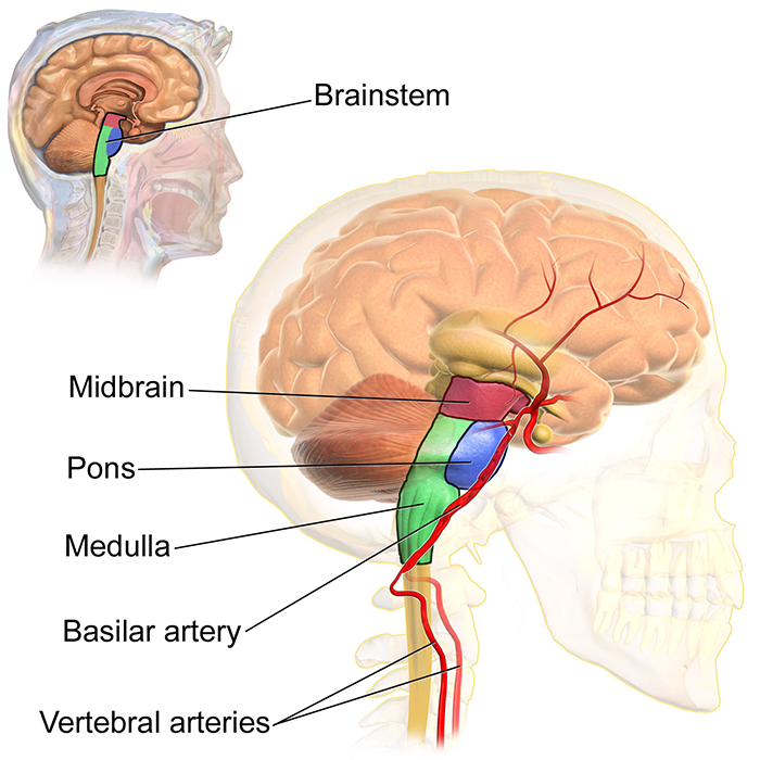
Located towards the base of your brain is a small but important region called the midbrain (derived from the developmental mesencephalon), which serves as a vital connection point between the other major regions of the brain - the forebrain and the hindbrain.
The midbrain is the topmost part of the brainstem, the connection central between the brain and the spinal cord. There are three main parts of the midbrain - the colliculi, the tegmentum, and the cerebral peduncles. Of the 12 cranial nerves, two thread directly from the midbrain - the oculomotor and trochlear nerves, responsible for eye and eyelid movement.
Colliculi
At the top of the midbrain are the colliculi, which derives its name from the Latin word for ‘hill. It contains two pairs of bulging, layered bundles of neurons called the superior and inferior colliculi. The superior ones work on preliminary processing of visual signals before they are passed on to the occipital lobe at the back of the head. The inferior ones do work on auditory signals before those are passed through the thalamus to the main auditory processing centre in the cortex.
Tegmentum
The tegmentum (Latin for ‘hood’) actually stretches down the length of the brainstem, but a portion of it forms a part of the midbrain. It contains two areas named after specific colours: the iron-rich red nucleus (which actually looks pink) is involved in the coordination of movements; the periaqueductal grey is a dense region of grey matter and is involved in suppressing pain. The tegmentum in the midbrain also contains connections that play a role in keeping us alert.
Cerebral peduncles
The back of the midbrain contains a pair of large nerve fibre bundles that connect the rest of the brainstem to the forebrain. These cerebral peduncles are the main highway for signals that need to be transported from the cortex to other parts of the central nervous system (CNS), and are especially important for body coordination.
Sandwiched between these bundles and the tegmentum is a layer called the substantia nigra, a darkly pigmented cluster of neurons with cells (containing melanin) that make the neurotransmitter dopamine; this layer of neurons is an important relay station for nerve signals of the CNS systems that coordinate our movements. This area is specifically damaged in Parkinson’s Disease.
Image credit: Wikimedia