The nervous system has units
Santiago Ramón y Cajal had a passion for art but followed his family into medicine. At the time, the prevailing theory was that the nervous system comprised one connected network of tissues. Using Camillo Golgi’s staining technique on microscopic slides of brain cells, Cajal saw through artistic eyes what others could not: that brain tissue was made of individual cells, called neurons. This was a major shift in knowledge of the brain at the time, and Golgi and Cajal were awarded a Nobel Prize for this discovery in 1906.
How neurons communicate
Sir Alan Hodgkin and Sir Andrew Huxley studied the neurons of squid, to show for the first time that signals travel along these nerve cells via electrical impulses from the movement of charged particles called ions. Sir John Eccles extended this work to explore what happens at the junction between two neurons, called a synapse. He showed that chemical signals can produce electrical currents that allow neurons to communicate. The trio was awarded the Nobel Prize in 1963 for these discoveries.
Shedding light on the visual brain
David Hubel and Torsten Wiesel advanced our understanding of vision. They showed different images to cats and noted which parts of their brains were activated. They devised a way to record the activity of a single neuron and then mapped out the visual cortex of the brain. They made discoveries about how the brain processes visual information. Importantly, they showed there's a critical period in the early stages of life when connections of the brain can be changed, and after that, visual pathways are set. They were awarded a Nobel Prize in 1981.
Roundworms: a simple model for the nervous system
Sydney Brenner was a geneticist whose contemporaries included the scientific luminaries James Watson and Francis Crick, who first described DNA’s structure as a double-stranded helix. Brenner, together with John Sulston and Bob Horvitz, turned their attention to mapping out the cells of a simple organism, the roundworm Caenorhabditis elegans. Their Nobel Prize–winning work opened up a new way for scientists to study neurons, of which roundworms have just 302. They showed that some neurons are programmed to die, and discovered the genes that regulate this process—which also happens in humans. This tiny worm can regenerate damaged neurons, providing a model to understand their growth and repair.
Where memory is stored in the brain
Only recently have scientists started to understand how we remember things and where memories are stored in the brain, at the level of neurons. Neuroscientist Eric Kandel was awarded the Nobel Prize for Physiology in 2000 for his work in showing that neurons change their connections when learning occurs. Kandel used a species of sea slug, Aplysia californica, as a simple model for studying what happens to neurons as an organism learns.
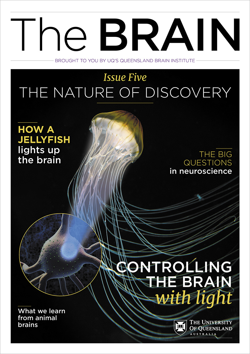
Lighting up the brain
Taking inspiration from nature, Professor Martin Chalfie wondered if the green glow of a jellyfish could be used to visualise cells in the body. Following the biochemical isolation of green fluorescent protein (GFP) by Osamu Shimomura, and mapping of its DNA sequence by Doug Prasher, Chalfie used GFP to visualise neurons in worms. Roger Tsien then developed many fluorescent proteins, which have been used in myriad ways, including to show how neurons grow and connect. Chalfie, Shimomura, and Tsien were recognised with the 2008 Nobel Prize in Chemistry for their transformational work.
Harnessing the gene editing power of bacteria
One of the most significant recent scientific developments has been the ability for scientists to easily target and modify DNA at specific places in the genome—known as gene editing. This game-changing technique, called CRISPR, came about when Professor Jennifer Doudna and Professor Emmanuelle Charpentier harnessed a natural defence that bacteria use to slice up and destroy viruses that invade them. Neuroscientists are using CRISPR to alter gene expression by editing DNA in mammalian brains under a variety of conditions. This may one day help us treat heritable brain disorders such as Huntington's and Alzheimer’s diseases, or to treat brain diseases caused by acquired DNA mutations, such as some brain cancers.
Human genome sequenced
In 2003, a 13-year project to map the whole human genome (3.2 billion base pairs of DNA) was completed. This key milestone in science couldn’t have happened without the development of Taq polymerase, an enzyme derived from heat-loving bacteria that were first found in Yellowstone National Park’s famous geysers. The mapping of 23,000 genes has enabled scientists to link particular genes with normal brain function, and find others that play a role in brain diseases and disorders.
Optogenetics - controlling the brain
Optogenetics is the process of using genetic techniques to enable light to turn cells like neurons on and off. This leap in neuroscience came initially from observing proteins in green algae that control movement in response to light. Scientists genetically engineer groups of neurons they want to study by inserting light-sensitive proteins called opsins. When light is shone on those neurons, they activate. This kind of stimulation is much more specific than using electrical stimulation, and can reveal how neurons connect and how they contribute to different behaviours.