Often in science, being curious and asking simple questions can lead to amazing discoveries. One such story began in the 1960s with a scientist, Osamu Shimomura, asking the question: what makes a jellyfish glow? Shimomura was able to identify the particular protein—which he called green fluorescent protein (GFP)—from the jellyfish Aequorea victoria that produced its bioluminescent light.
Years later, Professor Martin Chalfie heard about GFP and wondered if this protein could be used to highlight individual cells in other organisms. At the time, Professor Chalfie was using the tiny roundworm C. elegans to study how nerve cells worked. In a groundbreaking study, he successfully labelled groups of connected neurons in roundworms, and a new era in visualising nervous system cells began.
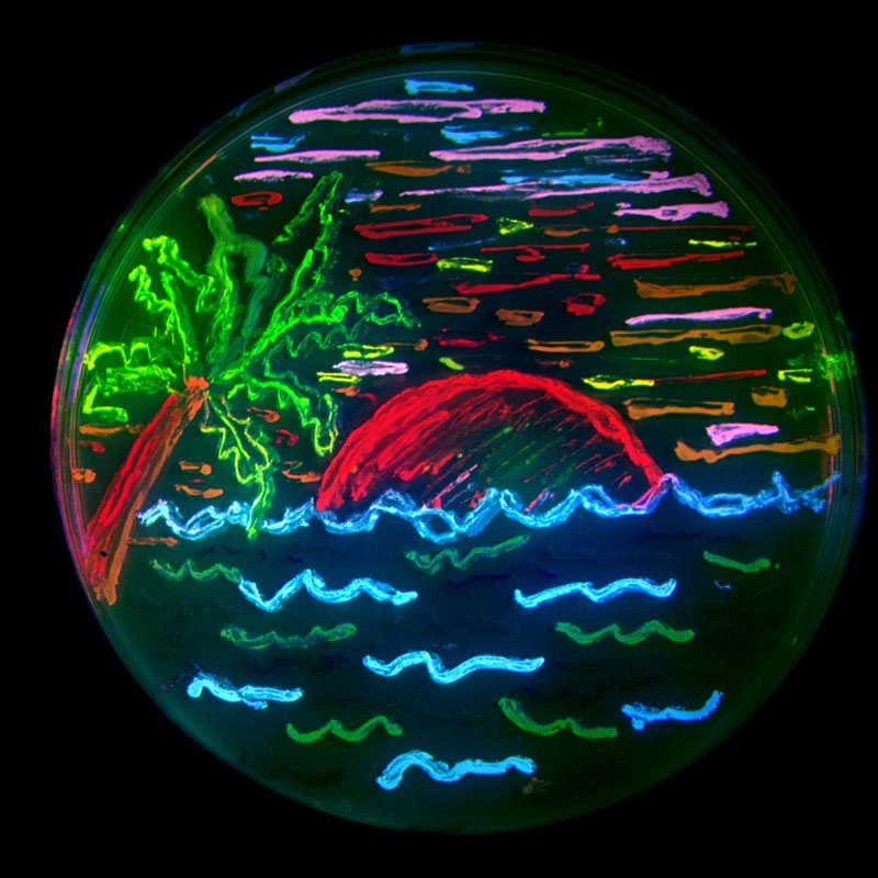
Scientists then used GFP to make all kinds of cells glow, from neurons to plant cells. But the glow didn’t last that long, and green wasn’t always the most ideal colour to work with. Enter biochemist Roger Tsien, who set about improving the brightness of GFP, as well as genetically engineering it to express more colours. He also isolated fluorescent proteins from other animals, to develop a broader range of colours. To showcase the array of colours, members of his team created a multicoloured beach scene with bacteria in a petri dish.
The use of GFP and other glowing proteins to light up the inner workings of organisms has revolutionised neuroscience. Before this, scientists weren’t able to see such detail.
Jellyfish protein lights up brain cells
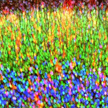
In brain research, GFP and other proteins have been used to visualise different neurons, to show how networks form in the brain, to identify individual neurons in slices of the brain, to study how neurons grow and connect, and to mark neurons affected by plaques that are hallmarks of Alzheimer’s disease. For their transformational work that began with a question about glowing jellyfish, all three scientists were recognised with the Nobel Prize in Chemistry in 2008.
Lighting up the tiniest parts of cells
One of the more exciting developments in neuroscience is using a fluorescent protein from the lobed cactus coral Lobophyllia hemprichii to highlight the workings inside a brain cell in real time.
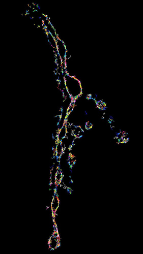
Using a highly sensitive super-resolution microscope to highlight important molecules that transmit signals at the junction (called a synapse) between neurons, Professor Frederic Meunier and his team at UQ’s Queensland Brain Institute have been able to reveal what happens inside living nerve cells.
“We can now see molecules organise themselves in real time, viewing the nitty gritty mechanisms that allow neurons to communicate,” says Professor Meunier.
“You see molecules moving randomly and then, for a few milliseconds, interacting with each other.”
“This is an exciting time, as we’ve opened the door for many more groundbreaking studies that will change our view of how molecules function to make the brain work. This work could further our understanding of memory, learning and neurodegenerative diseases,” he says.