Our brains are constantly receiving information from all around us via our senses, and generating movements that help us execute our goals. But our brains also have a rich repertoire of internally generated activity—our emotions, imaginings and memories—that isn’t triggered by what we just heard or saw, or the next movement we want to make. How does the brain achieve this? By what logic do those millions and millions of neurons work together to create a thought, or to convert these black squiggles into meaningful words and sentences, or to hit a cross-court forehand? This is a big question in neuroscience, and to answer it, scientists must track brain activity at the level of individual cells.
A new approach is to measure calcium, a key ion involved in a range of critical processes in the body, from contracting our muscles, to cell growth, memory formation, and sending signals along neurons. A change in the concentration of calcium ions is a way of tracking the activity of neurons. By tracking calcium, it is now possible to measure thousands of individual cells in animals as they execute specific actions.
Neuroscientists are using a cutting-edge technology called two-photon imaging microscopy to measure these changes. At UQ’s Queensland Brain Institute, Dr Lee Fletcher and his team use this technique to explore the way that dendrites, the fine branch-like extensions of neurons, process information in the brain. They have pinpointed that active dendritic information processing is important in brain circuits that are activated at the moment an animal is engaged in a task. The mammalian brain is so complex that to narrow down neurons from the approximately 70 million in a mouse brain, to just a single group—and link it to a behaviour—is an incredible advance.
Switching on brain cells at will
To go one step further than recording brain cell activity, Professor Williams and his team have used a revolutionary light-based technique called optogenetics to study how these circuits process information.
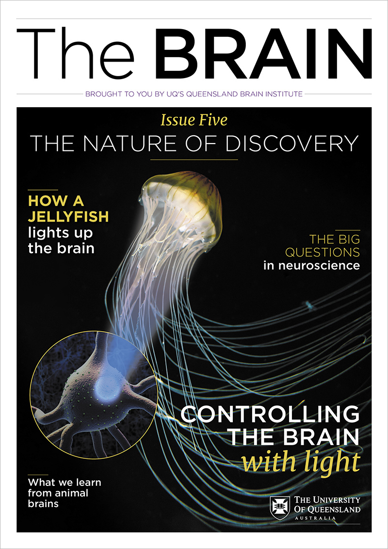
For this technique, scientists genetically engineer channels in a neuron to respond to light, by inserting a light-sensitive protein derived from algae. When a channel is exposed to light of a particular wavelength, say blue, it opens to let ions pass into the neuron to generate an electrical signal. Flashing a light can trigger or inhibit neurons expressing light-activated channels with amazing accuracy.
Using this technique, Professor Williams’ team has been able to identify a system of neurons that were active when animals were engaging in a task. This 'cholinergic system' is disrupted in diseases of the brain that impact cognitive abilities, such as dementia, so future research may reveal ways to stop this disruption and slow the progression of brain diseases.
Algae controls the brain

Who’d have thought that a humble slimy algae could progress the next revolution in neuroscience? In the 1990s, scientists observed that electrical signalling in cells of green algae changed in response to light. That kernel of knowledge was used to isolate the light-sensitive gene that has opened up a new field in neuroscience research, called optogenetics: using light to control the activity of brain cells in living tissue.