Vision
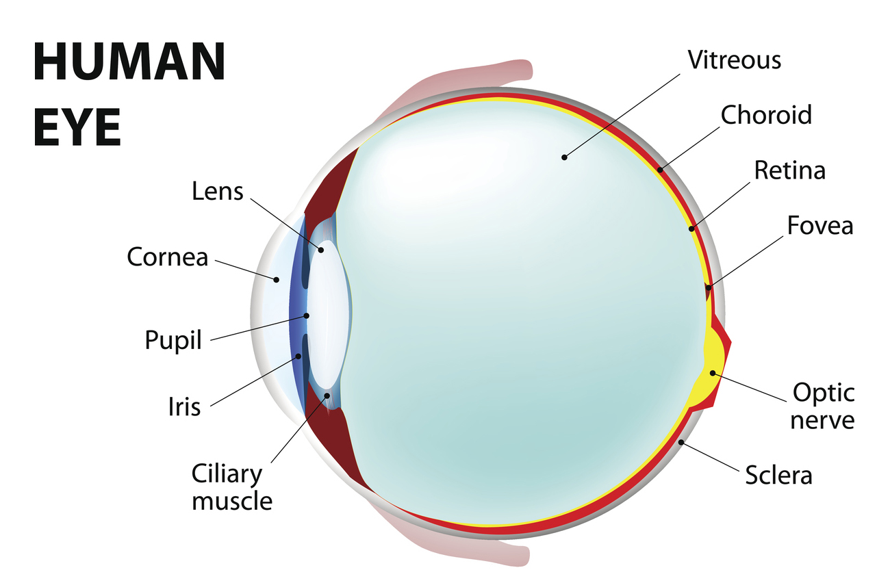
Let’s start with the eye. Light from the world around us enters the eye through the cornea, a curved structure that does much of the focusing work of the eye. It then passes through our pupil, whose diameter adjusts to control the amount of light allowed through, thanks to the contracting and dilating muscles of the iris. The light that passes through the pupil is then focused by the lens onto the retina, the first step in processing our visual world.
The retina
When light reaches our retina, it is detected by photoreceptors. Photoreceptors contain proteins that turn light energy into electrochemical signals, allowing cells in our nervous system to make sense of the visual world.
We have two broad types of photoreceptors, named for their shape: rods and cones. Rods are extremely sensitive to light and so are used only when light levels are low, like at night. Our rods are located mostly in peripheral regions of the retina, meaning our night-time vision is actually better the further from the centre of our field of view.
The less sensitive cones are used for daytime vision and are concentrated in the middle of our retina, particularly in a region called the fovea. The fovea is what gives us very sharp vision in the centre of our field of view.
We have three types of cones, sensitive to different wavelengths of light for the primary colours: red, green and blue. Called trichromatic theory, we see different colours of the visual spectrum when different proportions of the red, green and blue cells are activated.
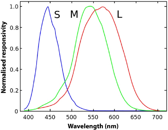
Vanessaezekowitz/Wikimedia
While humans have three types of cones for colour vision and can see all the colours of the rainbow, some species far outdo us. The mantis shrimp, for example, is a colourful crustacean found in tropical and subtropical waters, with 12 colour photoreceptors and the most complex visual system of any known animal. We don’t know what purpose this incredible biology, but it may serve to free up brain real estate for tasks other than distinguishing colours. You can find out more about the amazing visual system of mantis shrimp in the video below, courtesy of Professor Justin Marshall.
Why do we sometimes see afterimages?
One interesting consequence of how our colour vision works is the presence of afterimages. Afterimages are images that seem to remain, after looking away from another image. An example is included below. If you stare at the white dot in this image for at least 20 seconds then look to a blank white space, what do you see?

Instead of a blue star surrounded by a larger reddish border, you should see a faint yellow star surrounded by a greenish border: the yellow has replaced the blue, and the green has replaced the reddish colour.
This happens because we perceive yellow and blue, and green and red, as opponent colours. We can only see one of the opposing colours at a time because of the cells in the retina and visual cortex are activated by one colour and inhibited by another. We have three pairs of opponent colours: red-green, yellow-blue and black-white.
If we stare at an image like the one above, the photoreceptors producing the reddish signal become fatigued, reducing their ability to convert light to electrochemical signals. This means that less red signal reaches the brain, allowing the green-producing photoreceptor signal to dominate (such that the afterimage is green) until the other photoreceptors have recovered.
The building blocks of vision
After our photoreceptors have detected the light and converted the photons into electrochemical signals, the signals eventually make their way to ganglion cells. Ganglion cell axons then leave the retina and make their way to the brain via the optic nerve.
Between photoreceptors and ganglion cells, there are in fact a variety of other cell types that contribute to visual processing. These other cell types (such as bipolar cells, horizontal cells and amacrine cells) interact to give ganglion cells visual properties that form the building blocks of vision.
For example, some ganglion cells will respond best to a dark region surrounded by a ring of light; others prefer the opposite – a bright spot of light surrounded by a donut of darkness. These properties make ganglion cells very good at fundamental features of vision like detecting contrast. Other ganglion cells detect movement of light in a particular direction. This property, obviously, is useful for our ability to see objects that move across our field of vision.
To find out more about how cells of the retina interact to allow the direction of motion to be gauged, you can read this summary of work by QBI’s Professor Stephen Williams.