![]()
![]()
![]()
![]()
![]()
![]()
As the body's most complex and least-understood organ, the brain is as an object of much fascination and mistque, with many of its functions and structures still unknown. Weighing only 1.3kg - roughly 2% of the body's overall weight - not only does it perform incredibly diffcult and complex tasks, but it gives us our memories, our emotions, and defines who we are.
This year's neuroart entries from QBI researchers highlight this beauty, intricacy, and complexity through visuals. We share some of them with you here.
Competition winners
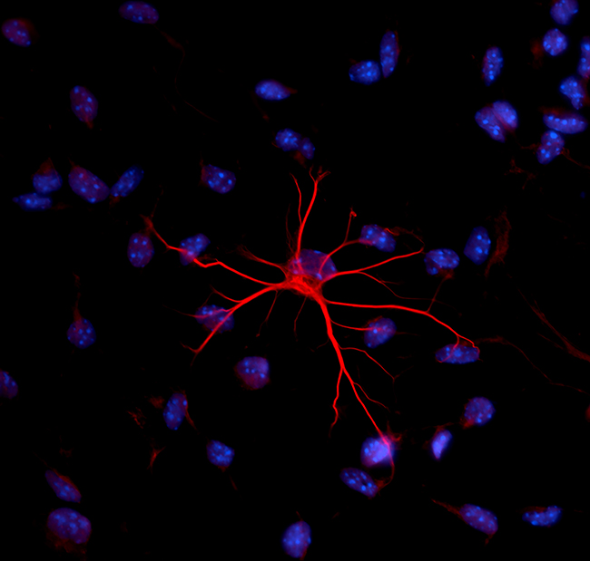
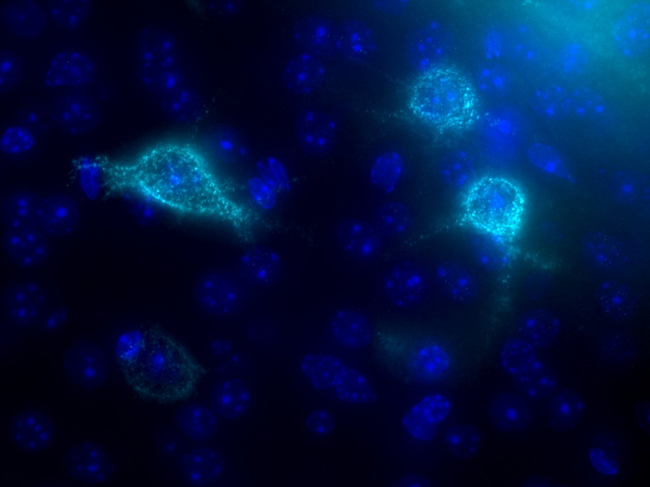
1st place: Jong Huat Tee, Postdoctoral research fellow, Medeiros lab, Outsanding brain's star.
A shining bright astrocyte that was isolated from a mouse's brain. A tool to study neuroinflammation in the Alzheimer's disease.
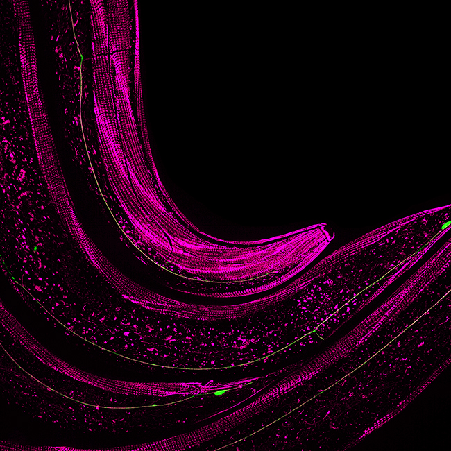
2nd place: Igor Bonacossa Pereira, PhD student, Hilliard lab, Heads or tails.
Here you see three worms smaller than a millimetre, with the sticky parts of its skin painted pink and its touch sensing neurons painted green.
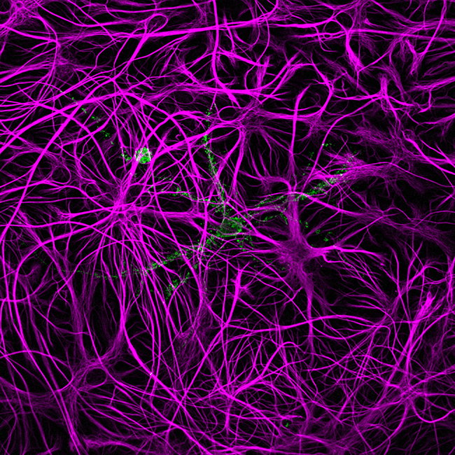
3nd place: Angelo Tedoldi, Postdoctoral research fellow, Sah lab, The haunted dish.
Glial cells are the most abundant cells within our brains and are extremely important for brain function. Here, we can see glial cells (magenta) haunting the petri dish and supporting the neuron (green) with filament like arms.
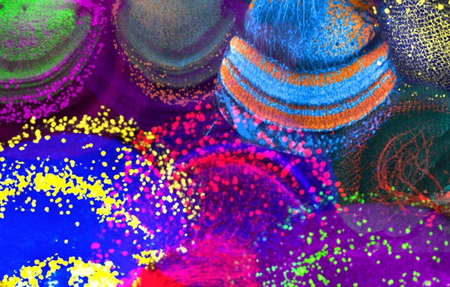
People's choice: Leonie Kirszenblat, Postdoctoral research fellow, van Swinderen lab, Intelligent life.
Inside the eye of a fruit fly there are many beautiful neurons that together create an alien landscape of which we know little about.
Finalists
Some other incredible images from the competition have been included below.
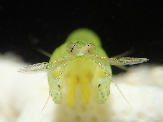
Hong Vo, PhD student, Marshall lab, Multiple points of view.
The stomatopod eye is divided into three regions, each containing its own pseudopupil. Together with the contralateral eye, the stomatopod is able to the view the world six times!
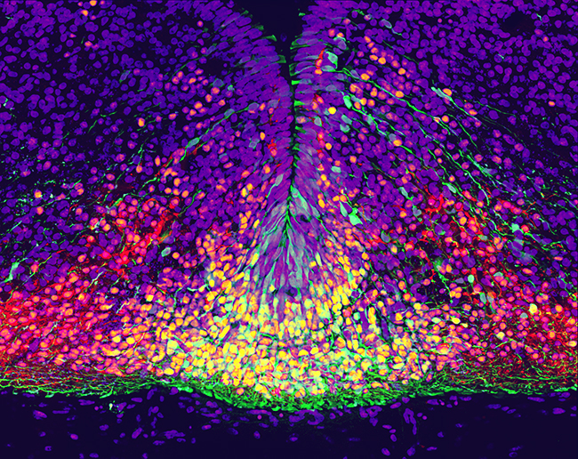
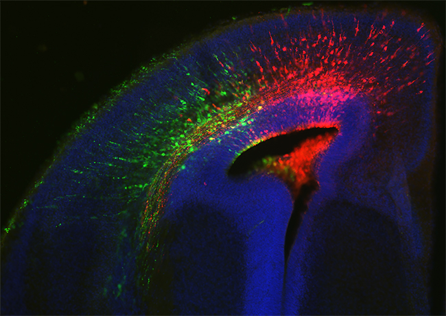
James Kesby, Postdoctoral research fellow, Eyles lab. Changing seasons.
Embryonic neuron development requires dynamic protein expression for maturation (yellow through red). The green cells were transfected with a fluorescent protein two days earlier and are migrating down and out to reach their final destination as mature dopamine neurons.
Phoebe Mayne, PhD student, Burne lab, Under the sea.
Since their discovery by Camillo Golgi in 1898, Perineuronal nets have been largely neglected by the scientific community. Albeit having a jellyfish-like appearance, perineuronal nets ensheath the soma, proximal dendrites and the axon segment of particular neurons. They are implicated in synaptic plasticity, regulating neuronal communication. Using fluorescence labelling, we were able to reveal just how detailed these structures are.
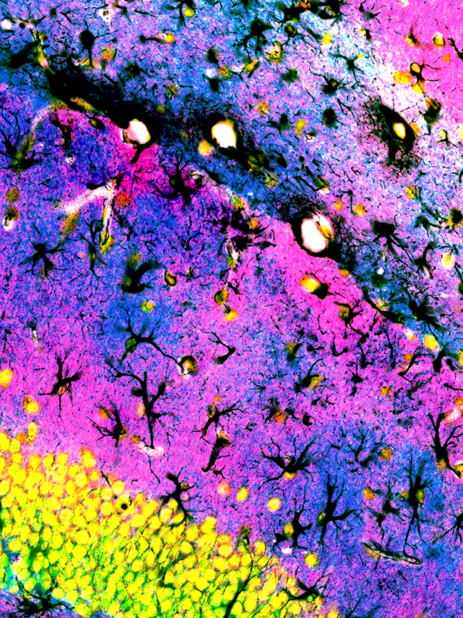
Robert Sullivan, Histology manager and postdoctoral researcher, Sah lab, Jacaranda and wattle blossoms.
Black branches and trunks of astrocytes holding up pink and blue islands of membrane transporters that regulate excitatory synaptic transmission of the yellow neurons.
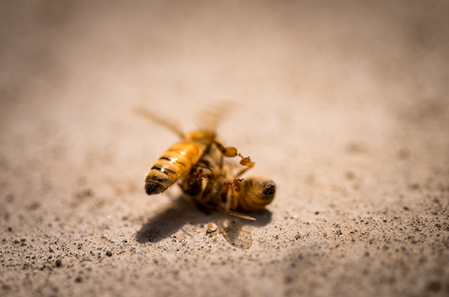
Kiaran Lawson, PhD student, Srinivasan lab, Bee-hind the scenes of a bee hive - the funeral director.
An undertaker bee takes off to carry a dead bee away from the hive. This demonstrates that bees are able to carry significant payloads even with their tiny actuators. Bees are studied at QBI to look at the science of flight and navigation, in order to create better robotic technology.
Lizzy Haines, PhD student, Richards lab, No, after you.
Neurons send out axons to connect with other brain regions. Two fluorescent labels are transfected to characterise which neuronal populations pioneer routes and which follow the lead.
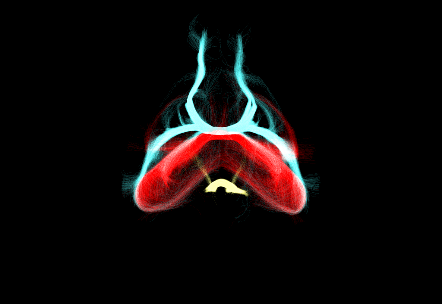
Yunan Ye, Honours student, Richards lab, Commisures.
The anterior (blue), hippocampal (red) and posterior (yellow) commissures connect the two brain hemispheres.
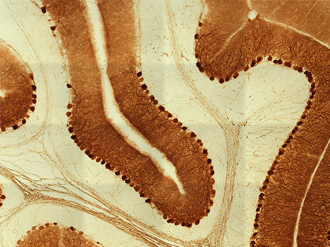
Asad Ali, Research assistant, Eyles lab, Purkinje cell layers.
These brilliant brown-stained Purkinje neurons, from within the cerebellum of an adult rat brain, are some of the largest and most complex cells in the mammalian brain. Each of these fascinating cells exhibits an abundance of very active long dendritic processes and are capable of receiving input from over 200,000 other cells in our brain.

")


