Stunning neuroscience images
![]()
![]()
![]()
![]()
![]()
![]()
![]()
QBI researchers produce beautiful images in the process of studying the brain. Here are just a few of them.
Find more images on our Instagram page @qldbraininstitute
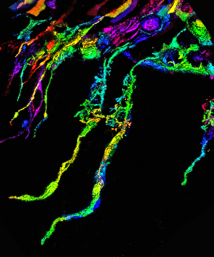
Blooming axons: this image comes from the hippocampus, a region of the brain important for learning and memory. Image by Iris Wang
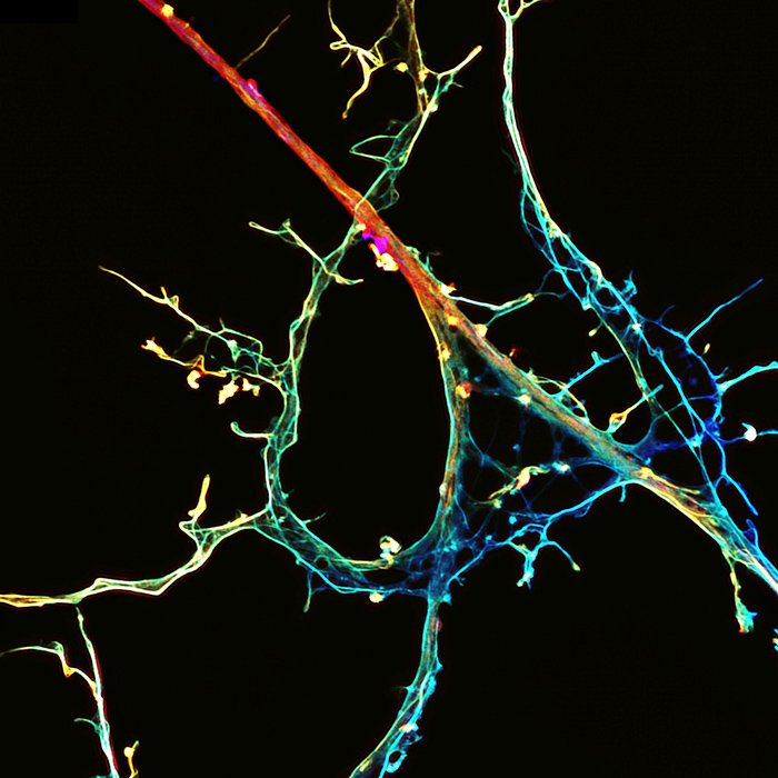
As neurons grow in culture they develop extensive networks and make connections with one another. The neuron shown here has been incubated with a protein, shown in pink, that binds to these synaptic regions. Image by Callista Harper.
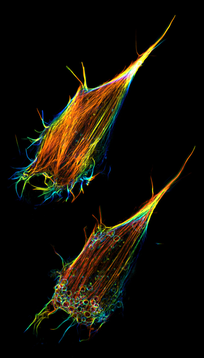
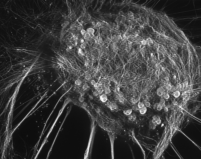
The actin cytoskeleton in neurosecretory cells undergoes a dramatic transformation after stimulation. The image displays a 3D reconstruction of the basal cortical actin network. The image was taken six minutes after stimulation with Ba2+, which causes secretion. In response, the cortical actin network undergoes remodelling. The acto-myosin II rings visible in the bottom left of the cell form to help recovering secretory vesicles that have fused with the plasma membrane. Image by Andreas Papadopulos, Meunier lab
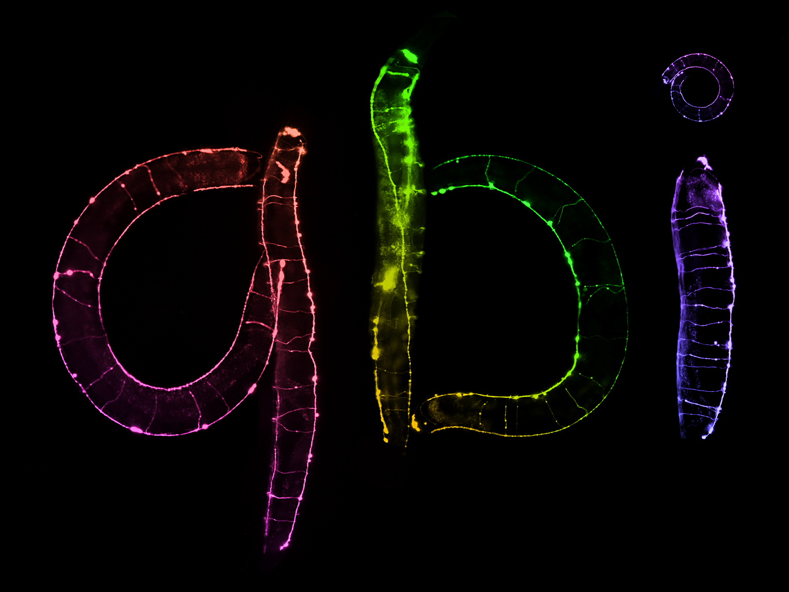
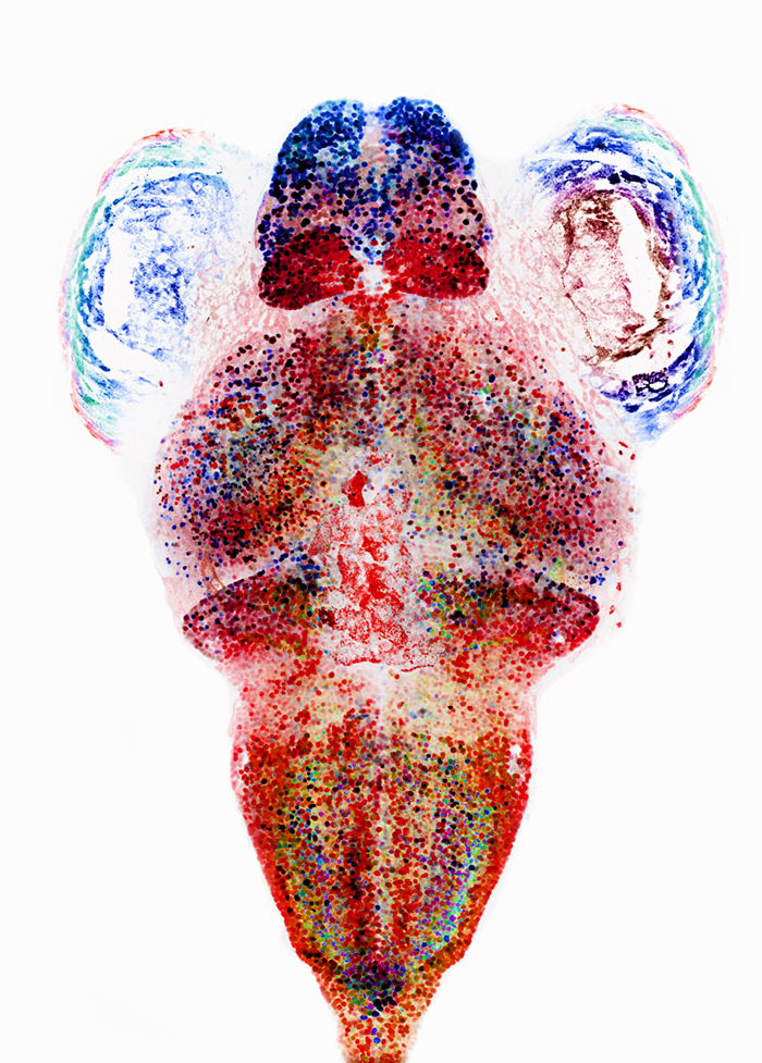
Part of QBI's core mission is to understand the fundamental processes of the brain. To do that, we often look to the simple roundworm (C. elegans). This worm is barely 1mm as an adult and has exactly 302 neurons. This simple system makes studying neurons much easier. Motor neurons of C. elegans innervate the entire worm's body, so when they are visualised with fluorescent proteins they show the profile of the animal, which have conveniently spelled out QBI! Image by Nick Valmas.
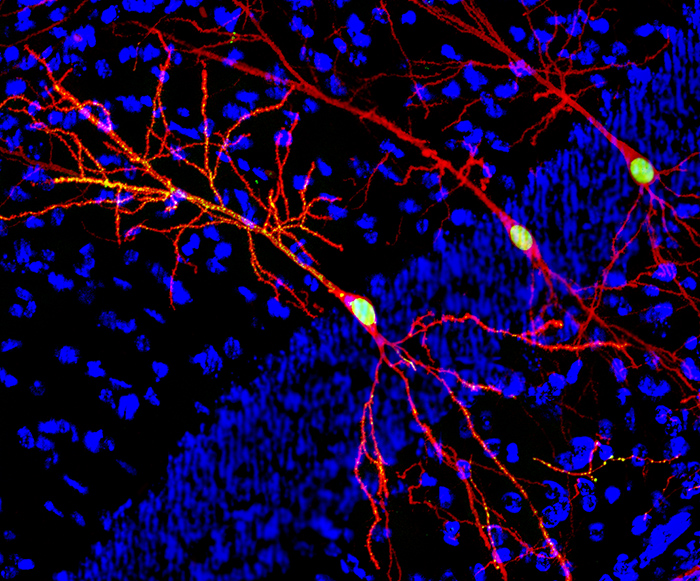
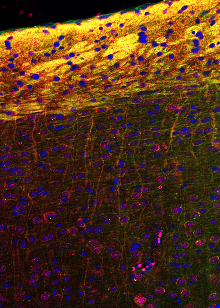
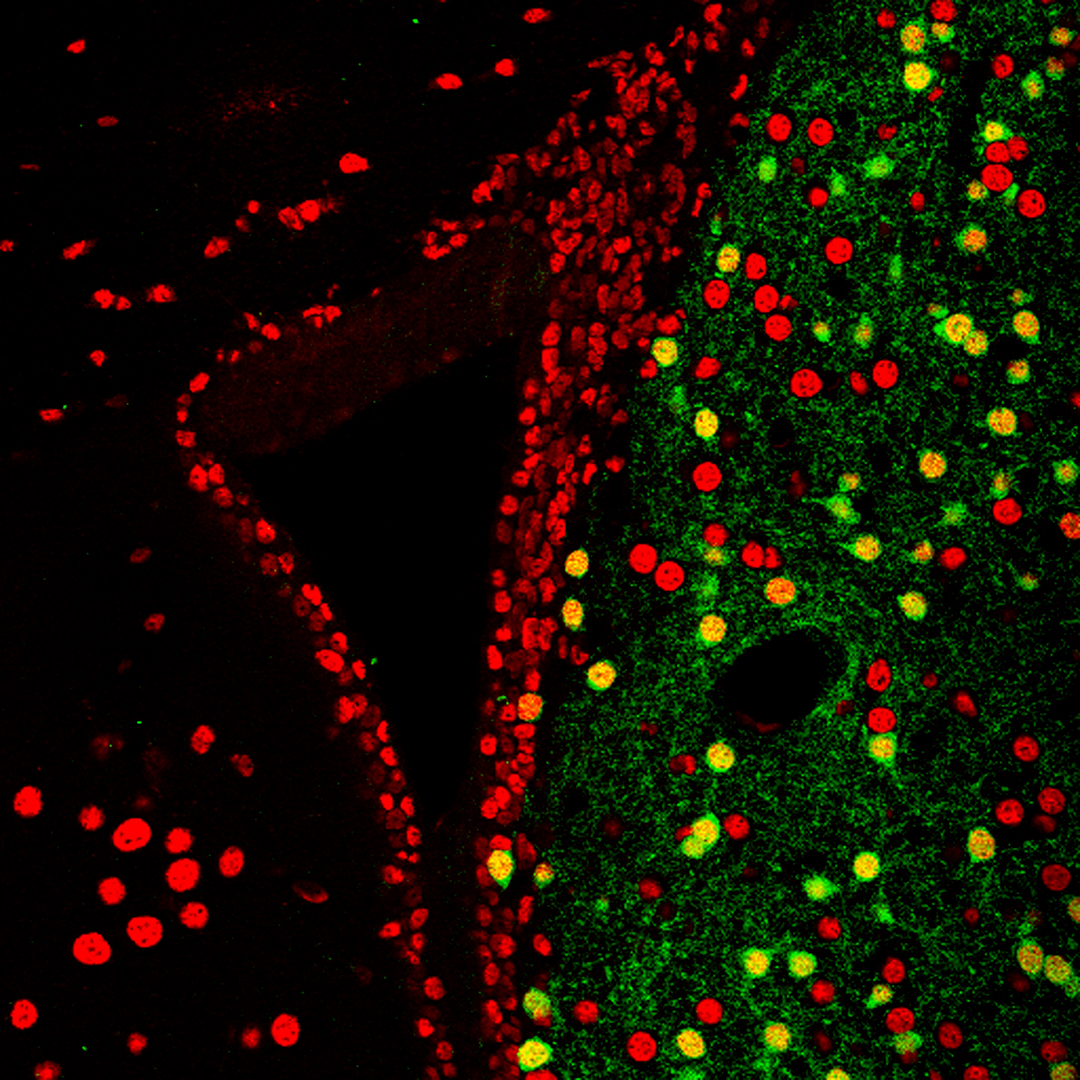
This is a histological section through the hippocampus with the cell bodies stained in blue. Neuron membranes are in red, and active presynaptic boutons and the cell nuclei in green. Image by Rodrigo Suarez.
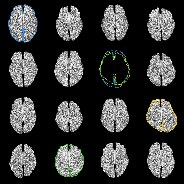
Our brains are amazing machines - and have quite an amount of variation between people. Here are scans of 15 brains from university students showing the similarities and differences in shape and folds. Image by Veronkia Halasz, former student in the Cunnington lab, which studies how the brain processes attention and predicting actions.
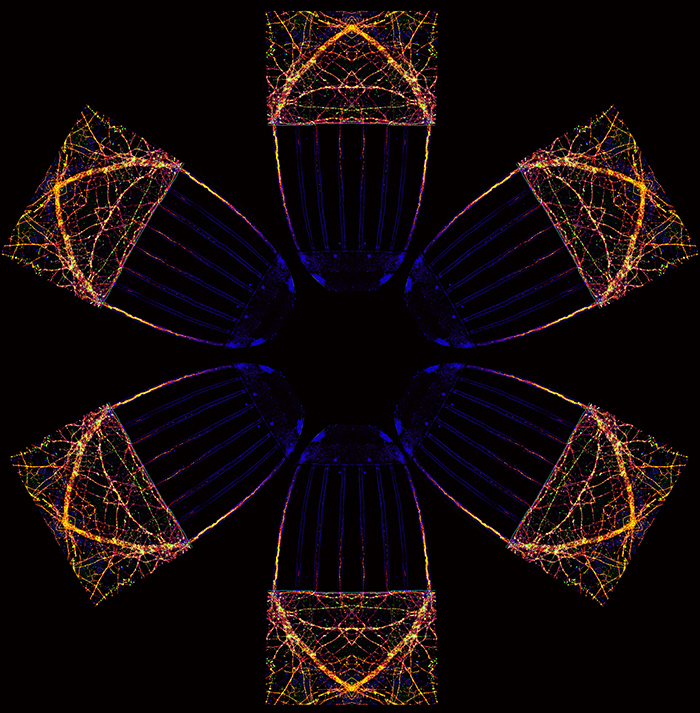
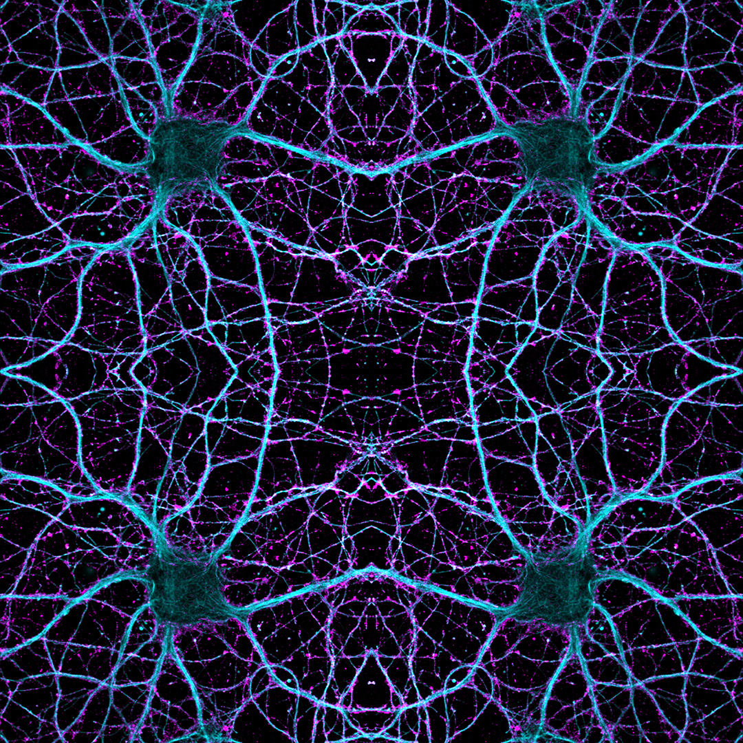
Axons (the cable of a neuron) from the hippocampus are finely separated (shown as the blue tracks) and labelled with colourful toxins (shown in green and red). Image by Dr Iris Wang in the Anggono lab, who is exploring neural plasticity and how receptors for glutamate are controlled in communication between neurons.
This tri-layered projection of a ganglion cell in the retina of the eye shows each of the neuron branches (dendrites) colour-coded according of their depth in the retina. (Red>Green>Blue). A recent study by Prof Williams showed that dendrites are not just passive structures for relaying signals, but are integral in processing information about moving light. Image by Ben Sivyer.
Aren't neurons beautiful! Cells in the first optic lobe (lamina) of mantis shrimps relay the information to the second (medulla) optic lobe. While humans see in 3 channels, mantis shrimp use at least 12, including UV and polarised light! They have the most-known complex visual system of any animal in the world. Image by Hanne Thoen from the Marshall lab, which researches the neuroscience of vision.
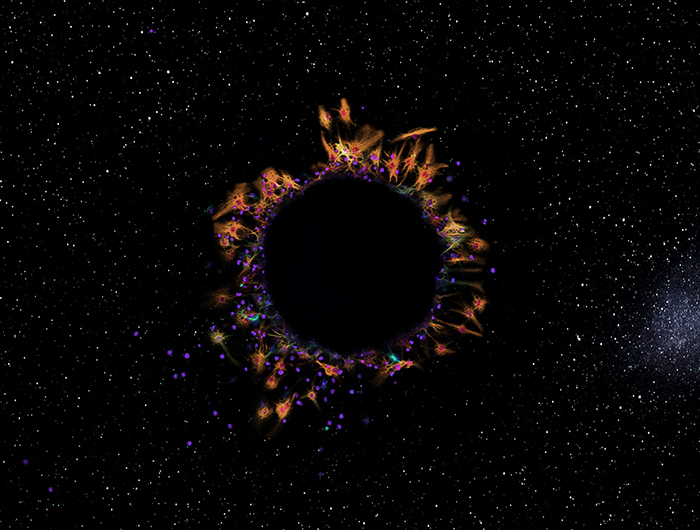
Single-plane image of a neurosphere, a ball of cells grown from a single stem cell. Glial cells (orange) and cell nuclei (purple) have migrated from the centre of the sphere. The occasional neuron (aqua) has also escaped the black centre of the sphere. Image: Chanel Taylor.
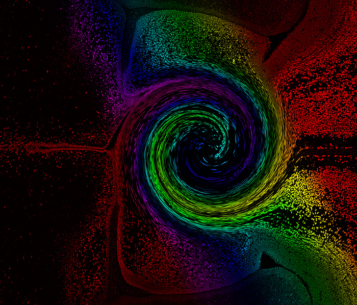
This swirling vortex-like image is a representation of the struggle brain cells endure. Cells from each side of the brain must travel a long way across the complex midline environment to make connections.
Image by Laura Morcom.
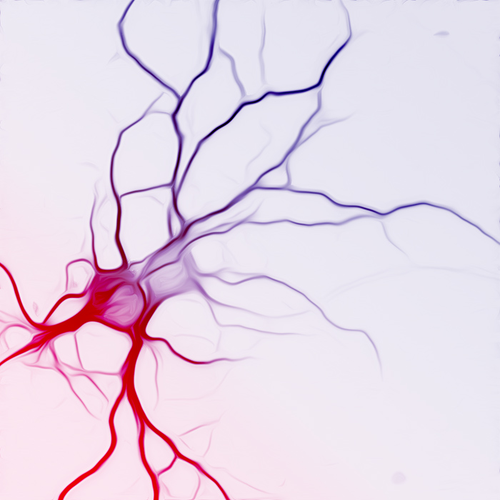
Two neurons interlace, forming connections and providing mutual support for growth in this beautiful, ethereal image. By Nadia Cummins.
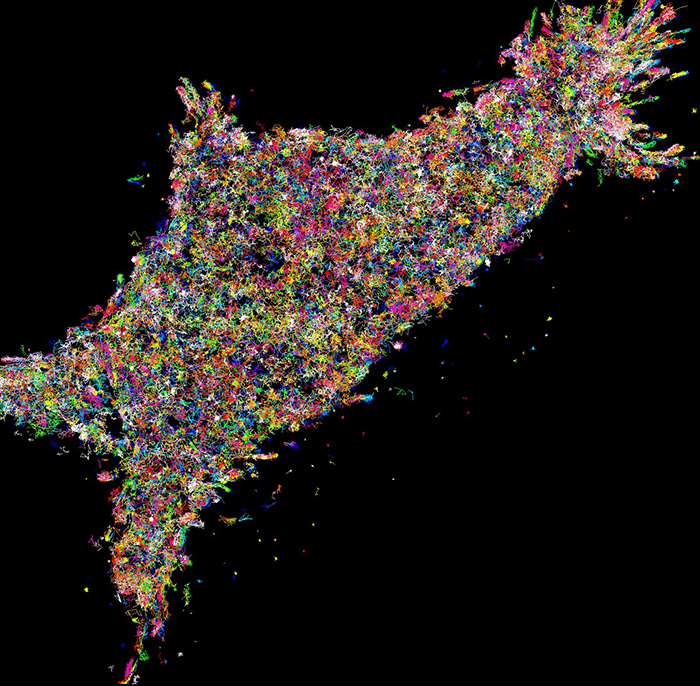
Where art meets science. A forest of neurons. Cortical (brain) neurons here are stained and magnified 20x. Image by Chanel Taylor.
This image shows the flight direction and wing patterns of the common budgie, when flying through a gap. Birds are ‘body aware’, and can fly through dense foliage with acute precision. Research from the Srinivasan Lab shows that budgies fly through openings wider than their wingspan without changing their wing position, but they close their wings when the opening is narrower (left). Image by Hong Vo and Ingo Schiffner.
This stunning image is a snapshot of a live tracking of the Munc18-1 loop mutant protein, which has been implicated in causes infant epilepsy and other neurodegenerative diseases. The Meunier lab looks at some of the smallest parts of the body - proteins a single molecule level - to track molecular processes of nerve cells and how they transmit messages. Image by Ravi Kiran Kasula.
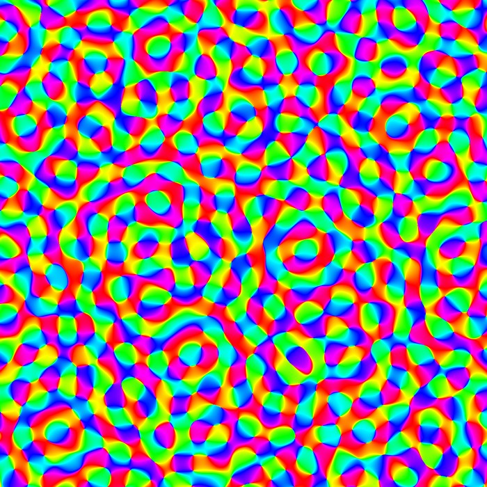
Are you mesmerised? This image is a simple stylised model showing the layout of the visual cortex of the brain. Image by J. Hunt.
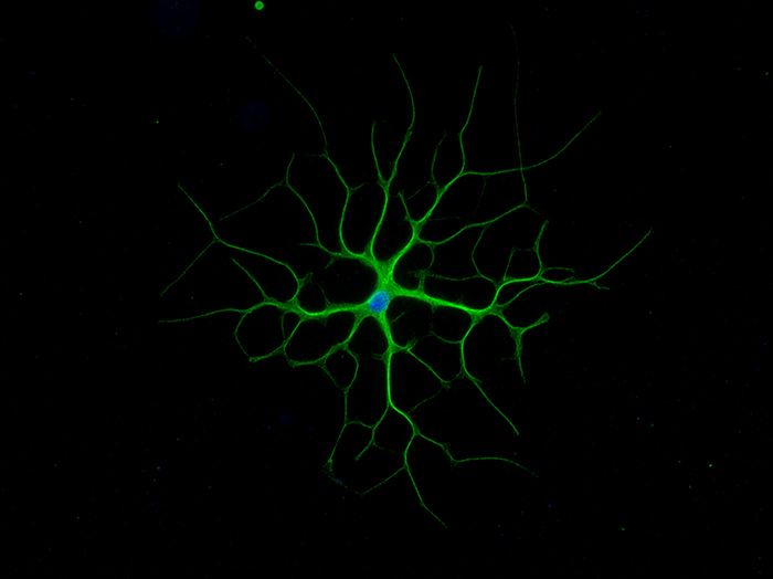
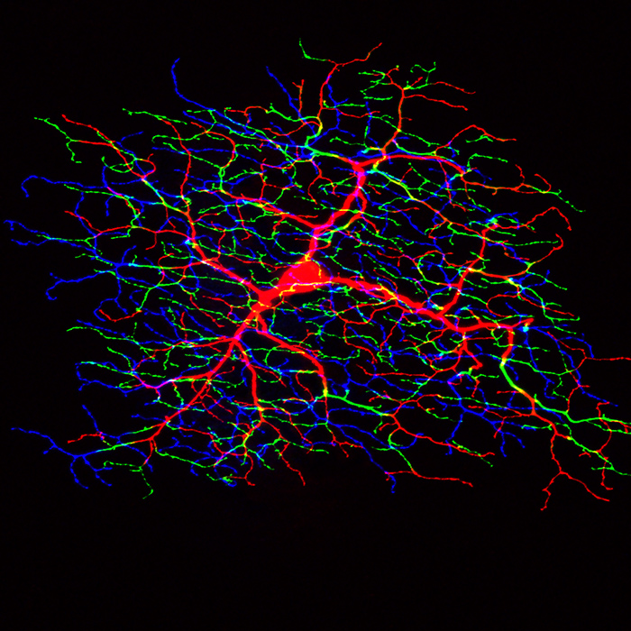
This beautiful star-shaped cell is an astrocyte. They are important for your nervous system as they maintain your neurons' working environment. Image by Dana Bradford.
An image of a zebrafish brain. This is a composite of over 5000 images (30GB) and reveals neurons at different depths within the brain shown in different colours. Image by Luke Hammond
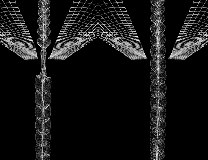
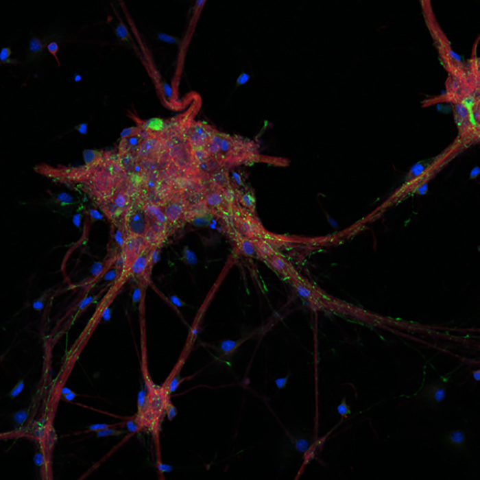
Reaching out: a stunning image of a dorsal root ganglion grown in a 3D collagen matrix. A dorsal root ganglion is is a cluster of nerve cell bodies in the spinal cord. Image by Zac Pujic.
Thunder and lightning: cells (blue) in the brain communicate through axons that share common pathways that look like thunder clouds (yellow). These axons project long distances to make electrical synapse that connected distal regions of the body. Image by Rob Sullivan
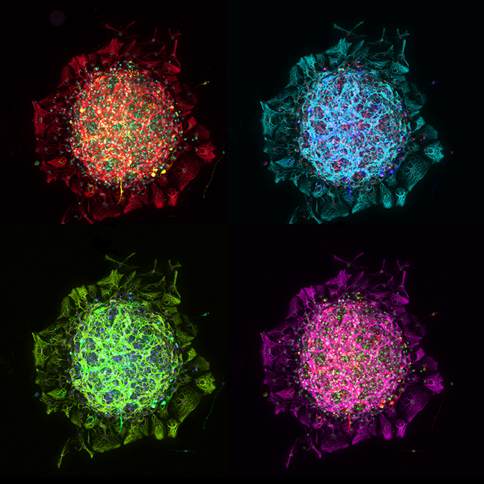
A differentiated neurosphere shown in four colours. Neurospheres are clumps of cells that form from a single stem cell—they give scientists a method to investigate neural precursors in the lab. Image: Chanel Taylor
Fantastical structures drive our fantastical imaginations. These are cultured cortical neurons, fluorescently labelled to visualise GABAergic inhibitory cells (green), L-type voltage gated calcium channels (red), and cell nuclei (blue), that arranged in a pattern reminiscent of Dahli’s spindly legged ‘Elephants’. Image by Helen Gooch.
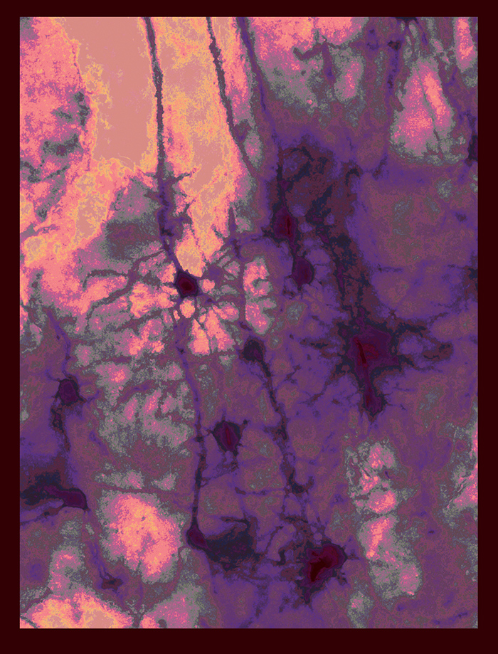
Nuclei in the brain have different shape, sizes and structures. They organise in layers and patterns that define motives in the tissue, resembling artwork. Image by J. Gonzalez.