In the ever-evolving world of cellular biology, the ability to observe proteins in action within their natural environment and at the nanoscale has long been a dream of neuroscientists such as Professor Fred Meunier and his team. Thanks to advances in super resolution microscopy, that dream is becoming a reality, accelerating what we understand about the inner dynamics of proteins in live cells.
In their latest paper, the Meunier lab introduces a powerful single-molecule super resolution imaging technique that opens exciting new avenues in cell biology, neuroscience, and drug discovery by revealing how proteins organise and signal in space and time at the nanoscale.
The new technique includes Fluorescent intrabody Localization Microscopy (FiLM) and a unique approach allowing nanoscale tracking of individual proteins in space and time as they change shape and location.
Professor Meunier explained that, unlike traditional single-particle tracking methods, FiLM uses nanobodies—tiny, specific antibody fragments— to target and visualise the cells’ own proteins.
“These unique nanobodies can distinguish between different shapes or states of the same protein, which offers us unprecedented insight into how these proteins behave, interact, and organise themselves into functional clusters,” Professor Meunier said.

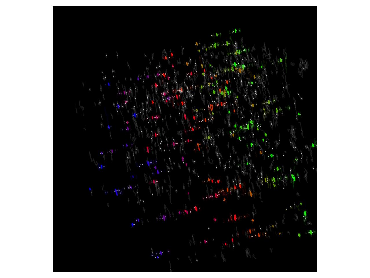
The protocol also introduces nanoscale spatiotemporal indexing clustering (NASTIC). This open-source tool extracts rich spatial and temporal data from single-molecule tracking.
NASTIC has a user-friendly interface and advanced clustering algorithms, which enable researchers to decode the protein’s complex choreography within live cells.
NASTIC’s architect and co-author in this study, Dr Tristan Wallis, explained that using this combined protocol, researchers can now see what was previously invisible.
“For the first time, we can monitor the behaviour of proteins, including those in specific functional states, in their native environment with both spatial and temporal precision,” Dr Rachel Gormal said, the lead author of this protocol.
“The analytical power of NASTIC allows us to study more comprehensively the nanoscale logic of the protein organisation, which underpins the brain’s signalling pathways, disease mechanisms and architecture.”
The Nature Protocol paper details how to perform nanoscale protein imaging and analysis for those with some experience in super-resolution microscopy and microscopy data analysis. The new protocol is compatible with cultured cells, primary neurons, and small model organisms such as C. elegans and zebrafish.


