
In big news for neuroscience, a team of American researchers recently mapped the human brain’s outler layer, the cerebral cortex, into 180 distinct regions.
Using imaging data from the Human Connectome Project – a United States government-led initiative to map the brain’s structural and functional connections – neuroscientists analysed the brains of 210 healthy young adults. The result was a modern atlas of the human brain, 97 areas of which have never been described before.
The cerebral cortex is the the folded outer layer that gives the brain its characteristic wrinkly appearance. It is divided into left and right hemispheres.
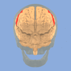
We know specific regions of the cortex are responsible for different roles. The primary somatosensory cortex, located on a vertical groove towards the middle of the brain, is the main area responsible for our sense of touch, for instance.
Most of what we understand about the detailed architecture of the brain comes from rodent studies. While brains of rats, mice and primates (us) are mostly similar in structure, they have distinct differences.
Unlike rodents, humans have a large prefrontal cortex, the area responsible for higher executive functions such as decision-making. We also communicate through language and as such have specific processing areas responsible for both creating speech and understanding it.
Improvements in techniques including functional magnetic resonance imaging (fMRI) – which measures brain activity by detecting blood flow changes – have enabled us to image living brains in real time in unprecedented detail.
The age-old neuroscience goal
Mapping the brain has been an aim for centuries, dating back to the pseudo-scientific discipline of phrenology in the 19th century, which posited that personality traits were located in specific parts of the brain.
Proponents would measure the skull over a corresponding brain area to determine, for example, how conscientious, benevolent or combative a person was.
More than a century ago, German anatomist Korbinian Brodmann classified the brain into specific areas based on the structure and organisation of cells in each region. Until now, this was the widely accepted map of brain regions, known as Brodmann’s areas.
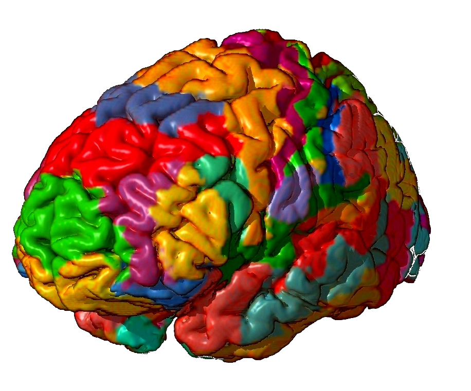
In the new study, researchers used a combination of different MRI images to map brain areas that are distinct in structure and function. They looked at physical structure, such as the thickness of the cortex, what areas were activated during certain tasks and whether this activity was coordinated with activity in other regions.
Some areas were predominantly associated with a single function, such as visual processing or movement. But many areas were not. In fact, scientists found networks of regions are activated even when the brain is in a resting state – when no explicit task is being performed.
A detailed brain map – so what?
The newly mapped brain is a landmark for neuroscience. An updated brain atlas will provide greater insights into how the brain controls behaviour and how disorders in certain regions contribute to brain diseases.
While rodent brain atlases originate from inbred strains of animals who vary little in their brain anatomy, individual variation in humans is common. There are differences between the anatomy of a person’s own left and right brain hemispheres, let alone the anatomical differences between individuals of different ages and genders.
For example, a recent study of 1,400 people found the left hippocampus, an area associated with memory, was usually larger in men than in women.
Because of this variation, it has historically been difficult to compare results from separate brain imaging studies and be certain the scans show activity in the same brain area. But now, the finer divisions of brain regions allow for better comparisons.
The brain map also has practical applications for neurosurgery. Currently, surgeons use a system of stereotaxic (3D) coordinates to determine and operate on specific brain regions. But this isn’t ideal as brain regions differ from person to person. The algorithm used to create the new atlas may now be used to personalise individual maps to help guide surgery more specifically.
Further categorisation
It’s likely the brain may be further compartmentalised into even more regions than the 180 already described. As imaging technology improves, we may discover further distinct sub-regions specialised in their makeup or activity.
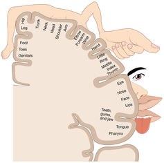
But the researchers also believe some of the newly mapped areas may later be found to be sub-areas, citing the primary somatosensory cortex as an example. This cortex is formed of what is called somatotopic sub-areas, which are brain regions that correspond point for point to sensory receptors at different parts of the body.
And different groups are beginning to map the genomic architecture of different brain regions. Together these new findings are going to lead to a detailed map of the whole of the human brain.


