Confocal Microscopes: Diskovery Spinning Disk Confocal (412)
An inverted spinning disk confocal microscope utilizing Spectral Applied Research’s Diskovery spinning disk module designed for both high-resolution and tissue imaging.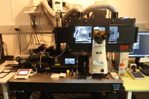
The Diskovery module runs on a Nikon TiE body and is controlled by Nikon NIS software.
This microscope is equipped with several high-end objectives suitable for imaging live cells and model organisms as well as fixed cells and tissues.
Features:
- Equipped with Diskovery disk head.
- 2x Zyla 4.2 sCMOS cameras enabling dual channel simultaneous imaging, fast capture of large mosaic images and high-sensitivity high dynamic range imaging.
- TIRF imaging capability
- 500µm wide range piezo z-drive and Nikon perfect focus for rapid z-stack capture and focus stabilisation.
Objectives:
Air:
- CFI Plan Apochromat Lambda 10x / N.A. 0.45 / W.D. 4.0mm
- CFI Plan Apochromat VC 20x / N.A. 0.75 / W.D. 1.00mm
Water:
- CFI Apo Lambda S LWD 40XWI / N.A. 1.15 / W.D. 0.60mm
Oil:
- CFI Apo Lambda 60x Oil / N.A. 1.4 / W.D. 0.13mm
- CFI Plan Apo Lambda 100x Oil / N.A. 1.45 / W.D. 0.13mm
- CFI Apochromat TIRF 60xH / N.A. 1.49 / W.D. 0.13mm
Lasers:
- 405nm, 488nm, 561nm, 640nm
Laser Power (Measurement at 10x (in mW)):
- 405nm (0.49mW)
- 488nm (1.63mW)
- 561nm (1.94mW)
- 640nm (1.65mW)
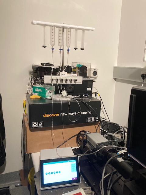
The Diskovery spinning disk confocal microscope has the Warner Instruments VCS-6 perfusion system, a computer-controlled valve control system that allows users to automate and control the delivery of solutions to imaging and recording chambers. For more information on the VCS-6, please visit the Warner Instruments page at:
https://www.warneronline.com/VCS-computer-controlled-Valve-Control-System
We use the VCS-6 perfusion system with the Warner Instruments DH-35iL culture dish incubator designed for glass-bottomed 35-mm dishes:
https://www.warneronline.com/culture-dish-incubator-dh-35il.
To cite use of the Diskovery in a paper please refer to the following suggestion (and adjust the objectives used as necessary):