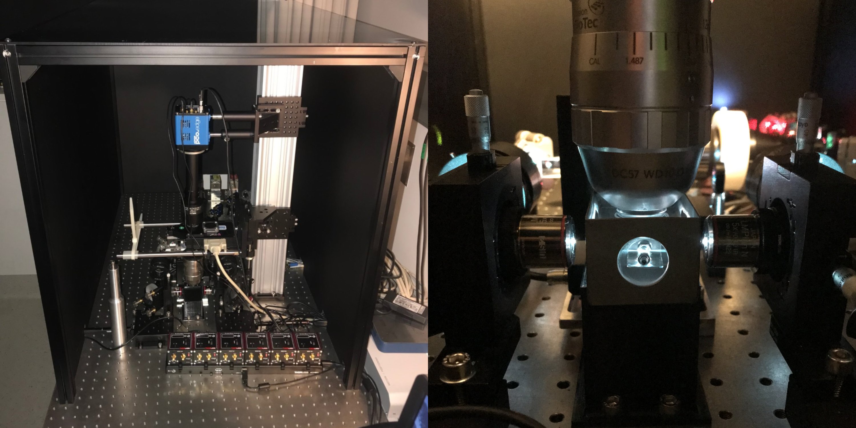
Cleared-Tissue Lightsheet Fluorescence Microscope (618)
QBI's custom-built cleared-tissue scanned light-sheet microscope uses scanning galvo mirrors (Cambridge Technology) to create excitation light sheets entering the sample from two opposite sides for double-sided illumination. These are focused using two Olympus 5x/0.1 NA objective lenses with a working distance of 23 mm. The fluorescence signal is detected orthogonally from the light-sheet plane by an 82 % QE pco.edge 4.2 sCMOS camera with Camera Link using the LaVision Biotec 12x/0.53 NA 8.5 mm WD MI PLAN objective for aqueous buffers (n=1.33-1.41), with dipping caps for DBE/BABB/DISCO/ECi (n=1.49-1.57, 10 mm WD) and CLARITY/CUBIC/ScaleS (n=1.42-1.48, 10.9 mm WD). Hardware integration is achieved by using custom-written LabVIEW software. Single-photon light-sheet illumination made possible by the incorporation of Coherent OBIS 488 nm, 561 nm and 640 nm lasers. Applications include volumetric imaging of cleared whole brains and organoids.
Features:
- 82 % QE pco.edge 4.2 sCMOS camer for excellent sensitivity and low noise imaging
- LaVision Biotec 12x/0.53 NA 8.5 mm WD MI PLAN objective
Objectives:
Clearing:
- LaVision Biotec 12x MultiImmersion / 0.53 NA / 8.5-10.9 mm WD / RI 1.33-1.57
- Olympus 10x MultiImmersion / 0.6 NA / 8 mm WD / RI 1.33-1.52
Fluorescent Filter Sets For:
- GFP / Alexa 488 / BP500-550
- Cy3 / Alexa 555/568 / BP570-640
- Cy5 / Alexa 647 / BP665-715
Lasers:
- 488nm, 561nm, 640nm 1-photon OBIS lasers
Book an instrument or training:
Contact
Microscopy Facility Manager
Rumelo Amor
r.amor@uq.edu.au
Senior Microscopy Officer
Andrew Thompson
a.thompson4@uq.edu.au
Histology Facility Manager
Rob Sullivan
qbi.histology@uq.edu.au
To contact the microscopy team please use:
qbi.microscopy@uq.edu.au